Abstract
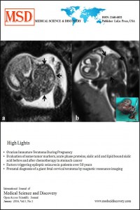
Fetal cervical teratomas are the rare forms of congenital teratomas with a high risk of perinatal morbidity and mortality. Imaging plays an essential role in the management of cervical teratoma and also helps in counselling parents. Ultrasound may be inadequate in the prenatal diagnosis of cervical teratoma due to large tumor size and fetal position. Magnetic resonance imaging could be useful in the in the work-up of tumours detected by ultrasound. We reported a 29-year old pregnant woman referred to our hospital with a finding of giant solid mass at the fetal neck. Ultrasound examination revealed a right-side mass sized 87x64x51 mm that extended from mandible to the anterior thoracic wall. Fetal magnetic resonance imaging provided additional information regarding exact anatomical location and extent of the mass. Thus, we found that fetal magnetic resonance imaging is a complementary diagnostic modality to antenatal ultrasound in the differential diagnosis of cervical teratoma.
References
- Tonni G, De Felice C, Centini G, Ginanneschi C. Cervical and oral teratoma in the fetus: a systematic review of etiology, pathology, diagnosis, treatment and prognosis. Arch Gynecol Obstet 2010; 282: 355-61.
- Thomas KA, Sohaey R. Congenital teratoma. Ultrasound Q 2012; 28: 197-9.
- Avni FE, Massez A, Cassart M. Tumors of the fetal body: a review. Pediatr Radiol 2009; 39: 1147-57.
- Araujo Júnior E, Guimarães Filho HA, Saito M, Pires AB, Pontes AL, Nardozza LM et al. Prenatal diagnosis of a large fetal cervical teratoma by three-dimensional ultrasonography: a case report. Arch Gynecol Obstet 2007; 275: 141-4.
- Silberman R, Mendelson IR. Teratoma of the neck: report of two cases and review of the literature. Arch Dis Child 1960; 35: 159-70.
- Trecet JC, Claramunt V, Larraz J, Ruiz E, Zuzuarregui M, Ugalde FJ. Prenatal ultrasound diagnosis of fetal teratoma of the neck. J Clin Ultrasound 1984; 12: 509-11.
- Jordan RB, Gauderer MW. Cervical teratomas: an analysis. Literature review and proposed classification. J Pediatr Surg 1988; 23: 583-91.
- Gorincour G, Dugougeat-Pilleul F, Bouvier R, Lorthois-Ninou S, Devonec S, Gaucherand P et al. Prenatal presentation of cervical congenital neuroblastoma. Prenat Diagn 2003; 23: 690-3.
- Figueiredo G, Pinto PS, Graham EM, Huisman TA. Congenital giant cervical teratoma: pre- and postnatal imaging. Fetal Diagn Ther 2010; 27: 231-2.
- MacArthur CJ. Prenatal diagnosis of fetal cervicofacial anomalies. Curr Opin Otolaryngol Head Neck Surg 2012; 20: 482-90.
- Rempen A, Feige A. Differential diagnosis of sonographically detected tumours in the fetal cervical region. Eur J Obstet Gynecol Reprod Biol. 1985; 20: 89-105.
- Nemec SF, Horcher E, Kasprian G, Brugger PC, Bettelheim D, Amann G et al. Tumor disease and associated congenital abnormalities on prenatal MRI. Eur J Radiol 2012; 81: 115-22.
- Hedrick HL. Ex utero intrapartum therapy. Semin Pediatr Surg 2003; 12:190-5.
Abstract
References
- Tonni G, De Felice C, Centini G, Ginanneschi C. Cervical and oral teratoma in the fetus: a systematic review of etiology, pathology, diagnosis, treatment and prognosis. Arch Gynecol Obstet 2010; 282: 355-61.
- Thomas KA, Sohaey R. Congenital teratoma. Ultrasound Q 2012; 28: 197-9.
- Avni FE, Massez A, Cassart M. Tumors of the fetal body: a review. Pediatr Radiol 2009; 39: 1147-57.
- Araujo Júnior E, Guimarães Filho HA, Saito M, Pires AB, Pontes AL, Nardozza LM et al. Prenatal diagnosis of a large fetal cervical teratoma by three-dimensional ultrasonography: a case report. Arch Gynecol Obstet 2007; 275: 141-4.
- Silberman R, Mendelson IR. Teratoma of the neck: report of two cases and review of the literature. Arch Dis Child 1960; 35: 159-70.
- Trecet JC, Claramunt V, Larraz J, Ruiz E, Zuzuarregui M, Ugalde FJ. Prenatal ultrasound diagnosis of fetal teratoma of the neck. J Clin Ultrasound 1984; 12: 509-11.
- Jordan RB, Gauderer MW. Cervical teratomas: an analysis. Literature review and proposed classification. J Pediatr Surg 1988; 23: 583-91.
- Gorincour G, Dugougeat-Pilleul F, Bouvier R, Lorthois-Ninou S, Devonec S, Gaucherand P et al. Prenatal presentation of cervical congenital neuroblastoma. Prenat Diagn 2003; 23: 690-3.
- Figueiredo G, Pinto PS, Graham EM, Huisman TA. Congenital giant cervical teratoma: pre- and postnatal imaging. Fetal Diagn Ther 2010; 27: 231-2.
- MacArthur CJ. Prenatal diagnosis of fetal cervicofacial anomalies. Curr Opin Otolaryngol Head Neck Surg 2012; 20: 482-90.
- Rempen A, Feige A. Differential diagnosis of sonographically detected tumours in the fetal cervical region. Eur J Obstet Gynecol Reprod Biol. 1985; 20: 89-105.
- Nemec SF, Horcher E, Kasprian G, Brugger PC, Bettelheim D, Amann G et al. Tumor disease and associated congenital abnormalities on prenatal MRI. Eur J Radiol 2012; 81: 115-22.
- Hedrick HL. Ex utero intrapartum therapy. Semin Pediatr Surg 2003; 12:190-5.
Details
| Primary Language | English |
|---|---|
| Journal Section | Case Reports |
| Authors | |
| Publication Date | January 15, 2016 |
| Published in Issue | Year 2016 Volume: 3 Issue: 1 |


