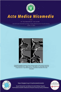
Uluslararası Frontal Sinüs Anatomi Sınıflamasına Göre Frontal Reses Hücre Prevalansının Bilgisayarlı Tomografik Analizi
Öz
Amaç: Sağlıklı paranazal sinüslerde frontal reses (FR) hücrelerinin prevalansını Uluslararası Frontal Sinüs Anatomisi Sınıflandırmasına (IFAC: International frontal sinus anatomy classification) dayalı olarak incelemek. Ayrıca, IFAC sisteminin gözlemciler arası uyumunu değerlendirmek.
Yöntem: Bu çalışmaya bilgisayarlı tomografik görüntülemelerinde (BT) paranazal sinüsleri hastalık bulunmayan 509 yetişkin hasta retrospektif olarak dahil edildi. İki araştırmacı birbirinden bağımsız olarak üç düzlemli BT rekonstrüksiyonlarını kullanarak 1018 taraftaki FR hücrelerini tanımladı. Her hücre tipinin prevalansı değerlendirildi ve gözlemciler arası uyum Kappa katsayısı (κ) kullanılarak ölçüldü.
Bulgular: Popülasyonumuzda, agger nasi hücresi (ANH) en yüksek prevalansa sahipti (%88,0), bunu supra bulla hücre (%43,0), supra agger hücre (SAH) (%25,0), frontal septal hücre (%22,0), supraorbital etmoid hücre (%17,1), supra agger frontal hücre (SAFH) (%8,3) ve supra bulla frontal hücre (SBFH) (%7,1) izledi. Bilateral insidans ANH için en yüksek (%80,4) ve SBFH için en düşük (%2,2) idi. SAH (kadınlarda %27,8; erkeklerde %22,2) ve SAFH (erkeklerde %11,6; kadınlarda %5,1) dışında diğer IFAC hücrelerinin prevalansı erkekler ve kadınlar arasında benzerdi. Vakaların %28,6'sında frontal sinüse pnömatize olan FR hücreleri gözlendi. Bu hücrelerin prevalansı erkek hastalarda kadınlara göre anlamlı derecede daha yüksekti. Tüm FR hücreleri için 0,94 ile 1,0 arasında değişen κ değerleri ile mükemmel bir gözlemciler arası uyum bulunmuştur.
Sonuç: FR hücrelerinin prevalansı popülasyona özgü farklılıklar göstermektedir. Cinsiyet farklılıkları, frontal sinüse pnömatize olan hücrelerin varlığını etkiler. IFAC, FR'deki hücreleri tanımlamak için güvenilir bir araçtır.
Anahtar Kelimeler: Uluslararası frontal sinüs anatomisi sınıflandırması, sinüs anatomisi, frontal hücreler, bilgisayarlı tomografi
Anahtar Kelimeler
Uluslararası frontal sinüs anatomisi sınıflandırması sinüs anatomisi frontal hücreler bilgisayarlı tomografi
Proje Numarası
KU-GOKAEK-2022/224
Kaynakça
- Wormald PJ, Hoseman W, Callejas C et al. The International Frontal Sinus Anatomy Classification (IFAC) and Classification of the Extent of Endoscopic Frontal Sinus Surgery (EFSS). Int Forum Allergy Rhinol.2016;6(7):677-696. doi:10.1002/alr.21738
- Valdes CJ, Bogado M, Samaha M. Causes of failure in endoscopic frontal sinus surgery in chronic rhinosinusitis patients. Int Forum Allergy Rhinol. 2014;4(6):502-506. doi:10.1002/alr.21307
- Van Alyea OE. Frontal Cells: An anatomic study of these cells with consideration of their clinical significance. Arch Otolaryngol. 1941;34(1):11-23. doi:10.1001/archotol.1941.00660040021003
- Bent JP 3rd, Spears RA, Kuhn FA, Stewart SM. Combined endoscopic intranasal and external frontal sinusotomy. Am J Rhinol. 1997;11:349-354. doi:10.2500/105065897781286098
- Kuhn FA. Chronic frontal sinusitis: the endoscopic frontal recess approach. Oper Tech Otolaryngol Head Neck Surg. 1996;7:222-229. doi:10.1016/S1043-1810(96)80037-6
- Lee WT, Kuhn FA, Citardi MJ. 3D computed tomographic analysis of frontal recess anatomy in patients without frontal sinusitis. Otolaryngol Head Neck Surg. 2004;131(3):164-173. doi:10.1016/j.otohns.2004.04.012
- Lund VJ, Stammberger H, Fokkens WJ et al. European position paper on the anatomical terminology of the internal nose and paranasal sinuses. Rhinol Suppl. 2014;24:1-34.
- Pianta L, Ferrari M, Schreiber A et al. Agger-bullar classification (ABC) of the frontal sinus drainage pathway: validation in a preclinical setting. Int Forum Allergy Rhinol. 2016;6:981-989. doi:10.1002/alr.21756
- Landis JR, Koch GG. The measurement of observer agreement for categorical data. Biometrics. 1977;1:159-174.
- Choby G, Thamboo A, Won TB, Kim J, Shih LC, Hwang PH. Computed tomography analysis of frontal cell prevalence according to the International Frontal Sinus Anatomy Classification. Int Forum Alergy Rhinol. 2018;8(7):825-830. doi:10.1002/alr.22105
- Gotlib T, Kołodziejczyk P, Kuźmińska M, Bobecka-Wesołowska K, Niemczyk K. Three-dimensional computed tomography analysis of frontoethmoidal cells: A critical evaluation of the International Frontal Sinus Anatomy Classification (IFAC). Clin Otolaryngol. 2019;44(6):954-960. doi:10.1111/coa.13412
- Pham HK, Tran TT, Nguyen TV, Thai TT. Multiplanar computed tomographic analysis of frontal cells according to International Frontal Sinus Anatomy Classification and their relation to frontal sinusitis. Reports in Medical Imaging. 2021;14:1-7. doi:10.2147/RMI.S291339
- Sjogren PP, Waghela R, Ashby S, Wiggins RH, Orlandi RR, Alt JA. International Frontal Sinus Anatomy Classification and anatomic predictors of low-lying anterior ethmoidal arteries. Am J Rhinol Allergy. 2017;1;31(3):174-176. doi:10.2500/ajra.2017.31.4428
- Tran LV, Ngo NH, Psaltis AJ. A radiological study assessing the prevalence of frontal recess cells and the most common frontal sinus drainage pathways. Am J Rhinol Allergy. 2019;33(3):323-330. doi:10.1177/1945892419826228
- Wormald PJ, Chan SZ. Surgical techniques for the removal of frontal recess cells obstructing the frontal ostium. Am J Rhinol. 2003;17(4):221-226.
- Marino MJ, Riley CA, Wu EL, Weinstein JE, Emerson N, McCoul ED. Variability of paranasal sinus pneumatization in the absence of sinus disease. Ochsner J. 2020;20(2):170-175. doi:10.31486/toj.19.0053
- Villarreal R, Wrobel BB, Macias-Valle LF et al. International assessment of inter- and intrarater reliability of the International Frontal Sinus Anatomy Classification system. Int Forum Allergy Rhinol. 2019;9(1):39-45. doi:10.1002/alr.22200
- Jaremek-Ochniak W, Sierdziński J, Popko-Zagor M. Three-dimensional computed tomography analysis of frontal recess cells according to the International Frontal Sinus Anatomy Classification (IFAC)-difficulties in identification of frontal recess cells in patients with diffuse primary chronic rhinosinusitis? Otolaryngol Pol. 2022;14;76(2):7-14. doi:10.5604/01.3001.0015.6959
Computed Tomographic Analysis of Frontal Recess Cell Prevalence According to International Frontal Sinus Anatomy Classification
Öz
Objective: To examine the prevalence of frontal recess (FR) cells based on the International Frontal Sinus Anatomy Classification (IFAC) in healthy sinuses, as well as evaluate the interrater agreement of the IFAC system.
Methods: Five hundred nine adult patients with non-diseased paranasal sinuses on computed tomography (CT) were retrospectively included in this study. Two researchers independently identified FR cells on 1018 sides using triplanar CT reconstructions. The prevalence of each cell type was assessed, and interobserver agreement was measured using the Kappa coefficient (κ).
Results: In our population, the agger nasi cell (ANC) had the highest prevalence (88.0%), followed by supra bulla cell (43.0%), supra agger cell (SAC) (25.0%), frontal septal cell (22.0%), supraorbital ethmoid cell (17.1%), supra agger frontal cell (SAFC) (8.3%), and supra bulla frontal cell (SBFC) (7.1%). Bilateral incidence was highest for the ANC (80.4%) and lowest for the SBFC (2.2%). The prevalence of most IFAC cells was similar between males and females, except in SAC (27.8% in females vs. 22.2% in males) and SAFC (11.6% in males vs. 5.1% in females). FR cells that pneumatize into the frontal sinus were observed in 28.6% of cases, with a significantly higher prevalence in male patients compared to females. Excellent interrater agreement was found for all FR cells, with κ values ranging from 0.94 to 1.0.
Conclusion: The prevalence of FR cells demonstrates variations specific to the population. Gender differences appear to influence the presence of cells pneumatizing into the frontal sinus. The IFAC is a reliable tool for identifying cells in the FR.
Keywords: International frontal sinus anatomy classification, sinus anatomy, frontal cells, computed tomograpy
Anahtar Kelimeler
International frontal sinus anatomy classification sinus anatomy frontal cells computed tomograpy
Etik Beyan
Approval was granted by the Ethics Committee of the Kocaeli University Faculty of Medicine (KU-GOKAEK-2022/224).
Destekleyen Kurum
none
Proje Numarası
KU-GOKAEK-2022/224
Teşekkür
We are grateful to Assistant Professor Sibel Balcı for assisting us in the statistical analysis.
Kaynakça
- Wormald PJ, Hoseman W, Callejas C et al. The International Frontal Sinus Anatomy Classification (IFAC) and Classification of the Extent of Endoscopic Frontal Sinus Surgery (EFSS). Int Forum Allergy Rhinol.2016;6(7):677-696. doi:10.1002/alr.21738
- Valdes CJ, Bogado M, Samaha M. Causes of failure in endoscopic frontal sinus surgery in chronic rhinosinusitis patients. Int Forum Allergy Rhinol. 2014;4(6):502-506. doi:10.1002/alr.21307
- Van Alyea OE. Frontal Cells: An anatomic study of these cells with consideration of their clinical significance. Arch Otolaryngol. 1941;34(1):11-23. doi:10.1001/archotol.1941.00660040021003
- Bent JP 3rd, Spears RA, Kuhn FA, Stewart SM. Combined endoscopic intranasal and external frontal sinusotomy. Am J Rhinol. 1997;11:349-354. doi:10.2500/105065897781286098
- Kuhn FA. Chronic frontal sinusitis: the endoscopic frontal recess approach. Oper Tech Otolaryngol Head Neck Surg. 1996;7:222-229. doi:10.1016/S1043-1810(96)80037-6
- Lee WT, Kuhn FA, Citardi MJ. 3D computed tomographic analysis of frontal recess anatomy in patients without frontal sinusitis. Otolaryngol Head Neck Surg. 2004;131(3):164-173. doi:10.1016/j.otohns.2004.04.012
- Lund VJ, Stammberger H, Fokkens WJ et al. European position paper on the anatomical terminology of the internal nose and paranasal sinuses. Rhinol Suppl. 2014;24:1-34.
- Pianta L, Ferrari M, Schreiber A et al. Agger-bullar classification (ABC) of the frontal sinus drainage pathway: validation in a preclinical setting. Int Forum Allergy Rhinol. 2016;6:981-989. doi:10.1002/alr.21756
- Landis JR, Koch GG. The measurement of observer agreement for categorical data. Biometrics. 1977;1:159-174.
- Choby G, Thamboo A, Won TB, Kim J, Shih LC, Hwang PH. Computed tomography analysis of frontal cell prevalence according to the International Frontal Sinus Anatomy Classification. Int Forum Alergy Rhinol. 2018;8(7):825-830. doi:10.1002/alr.22105
- Gotlib T, Kołodziejczyk P, Kuźmińska M, Bobecka-Wesołowska K, Niemczyk K. Three-dimensional computed tomography analysis of frontoethmoidal cells: A critical evaluation of the International Frontal Sinus Anatomy Classification (IFAC). Clin Otolaryngol. 2019;44(6):954-960. doi:10.1111/coa.13412
- Pham HK, Tran TT, Nguyen TV, Thai TT. Multiplanar computed tomographic analysis of frontal cells according to International Frontal Sinus Anatomy Classification and their relation to frontal sinusitis. Reports in Medical Imaging. 2021;14:1-7. doi:10.2147/RMI.S291339
- Sjogren PP, Waghela R, Ashby S, Wiggins RH, Orlandi RR, Alt JA. International Frontal Sinus Anatomy Classification and anatomic predictors of low-lying anterior ethmoidal arteries. Am J Rhinol Allergy. 2017;1;31(3):174-176. doi:10.2500/ajra.2017.31.4428
- Tran LV, Ngo NH, Psaltis AJ. A radiological study assessing the prevalence of frontal recess cells and the most common frontal sinus drainage pathways. Am J Rhinol Allergy. 2019;33(3):323-330. doi:10.1177/1945892419826228
- Wormald PJ, Chan SZ. Surgical techniques for the removal of frontal recess cells obstructing the frontal ostium. Am J Rhinol. 2003;17(4):221-226.
- Marino MJ, Riley CA, Wu EL, Weinstein JE, Emerson N, McCoul ED. Variability of paranasal sinus pneumatization in the absence of sinus disease. Ochsner J. 2020;20(2):170-175. doi:10.31486/toj.19.0053
- Villarreal R, Wrobel BB, Macias-Valle LF et al. International assessment of inter- and intrarater reliability of the International Frontal Sinus Anatomy Classification system. Int Forum Allergy Rhinol. 2019;9(1):39-45. doi:10.1002/alr.22200
- Jaremek-Ochniak W, Sierdziński J, Popko-Zagor M. Three-dimensional computed tomography analysis of frontal recess cells according to the International Frontal Sinus Anatomy Classification (IFAC)-difficulties in identification of frontal recess cells in patients with diffuse primary chronic rhinosinusitis? Otolaryngol Pol. 2022;14;76(2):7-14. doi:10.5604/01.3001.0015.6959
Ayrıntılar
| Birincil Dil | İngilizce |
|---|---|
| Konular | Kulak Burun Boğaz |
| Bölüm | Araştırma Makaleleri |
| Yazarlar | |
| Proje Numarası | KU-GOKAEK-2022/224 |
| Yayımlanma Tarihi | 30 Haziran 2024 |
| Gönderilme Tarihi | 3 Mart 2024 |
| Kabul Tarihi | 28 Haziran 2024 |
| Yayımlandığı Sayı | Yıl 2024 Cilt: 7 Sayı: 2 |
"Acta Medica Nicomedia" Tıp dergisinde https://dergipark.org.tr/tr/pub/actamednicomedia adresinden yayımlanan makaleler açık erişime sahip olup Creative Commons Atıf-AynıLisanslaPaylaş 4.0 Uluslararası Lisansı (CC BY SA 4.0) ile lisanslanmıştır.


